Dolphin Ceph Tracing

Dolphin Imaging software is an FDA-cleared Class II medical device

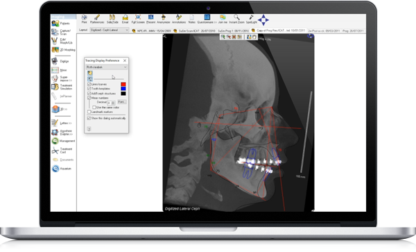
Ceph Tracing allows you to analyze cephalometric radiographs and create progress superimpositions quickly and accurately. Utilized by thousands of private practices throughout the world and most orthodontic and oral surgery training programs in North America, Dolphin Ceph Tracing software dramatically reduces the tedious and time-consuming task of cephalometric tracing.

Supports a Variety of Input

  • Capture images from existing files, flatbed scanner, other TWAIN sources or digital x-ray systems. Or, create 2D radiographs out of volumetric CBCT data, using optional Dolphin 3D software.

Lateral Cephalometric Analysis for Orthodontic and Surgical Use

  • Lateral analyses include Ricketts, McNamara, Steiner (Tweed), Jarabak, Roth, Sassouni, McLaughlin, Downs-Northwestern, Bjork, Alexander (Vari-Simplex), Holdaway, Alabama, Burstone, Gerety and many other variations; more than 400 in all!
  • Frontal analyses include Ricketts, Van Arsdale, Grummons and Grummons Simplified.
  • Arch analysis: Bolton, Schwarz.
  • Automated tooth and cephalometric structure templates, including soft tissue profile, airway, symphysis, maxilla, mandible, condyle, developing third molar, inferior alveolar canal, mastoid process, nasal cavities, orbits, zygomatic arch and many more.
  • Professional presentation: measurement table, tracing superimposition, tracing and image.
  • Norm deviation display “Downs Wiggle Gram.”
  • Fully customizable cephalometric analysis, structure, norms.
  • Norms categorized by gender, age, race.

Superimpositions

  • Easily overlay tracings over the patient’s lateral photograph or radiographic images.
  • Superimpose tracings of different timepoints with standardized superimposition references such as SN at Sella, Frankfort at Porion, Na-Pg at ANS-PNS, Na-Ba at CC, Na-Ba at Na, ANS-PNS at ANS, Go-Me at Me.
  • Multiple superimpositions.
  • Free-form superimposition.
  • 5-part Ricketts superimposition.
  • Calculate an “average tracing” from a sample group of patients for research purposes.
  • Create default settings from your current tracing superimpositions.

Digitizing Procedure

  • Simple x-ray calibration.
  • Illustrations and detailed description, depicting each landmark location.
  • Automatically generated anatomical structures and profiles.
  • Image enhancements: zoom in/out, brightness, contrast, sharpness, emboss.
  • Tracing alignment: Frankfort Horizontal, Horizontal Plane, Natural Head Posture.

Other Features

  • Powerful Image Enhancement features to uncover hard-to-see structures.
  • Integration with Microsoft PowerPoint and Microsoft Excel.
  • Overlaying tracing from different timepoints while digitizing—ensuring the most accurate superimposition.

Requirements - Specifications