Dolphin 3D Surgery™

Dolphin Imaging software is an FDA-cleared Class II medical device

What our customers say:

“My patients are more and more asking to pre-visualize their face ‘with’ or ‘without‘ surgery and choose their treatment accordingly. Dolphin 3D Surgery is a wonderful tool of communication between doctor team and patient. It allows the patients to push away the fear of surgery.”
– Dr. Alain Souchet, Mulhouse, France, customer since 2004

“Since my involvement with computed imaging research in the early 1980’s, I have been waiting for user-friendly 3D orthognathic planning software. Thirty years later, now we have it. Dolphin 3D Surgery is user-friendly and builds on the intuitive 2D planning interface.”
– Dr. Paul M. Thomas, Chapel Hill, NC, customer since 1995

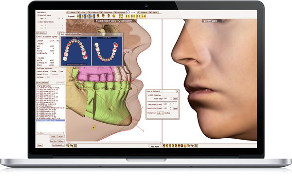
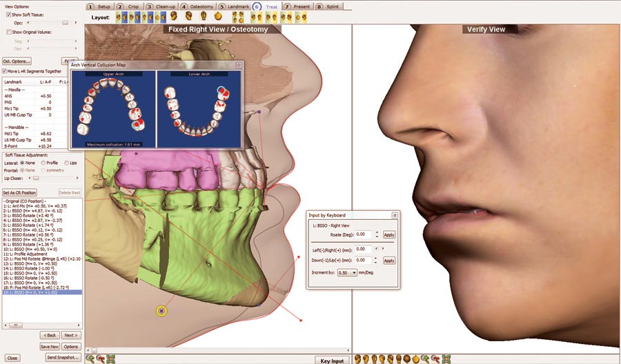
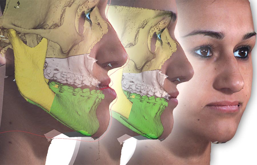
Interactive, Instantaneous, Incredible…

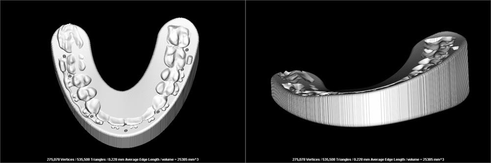
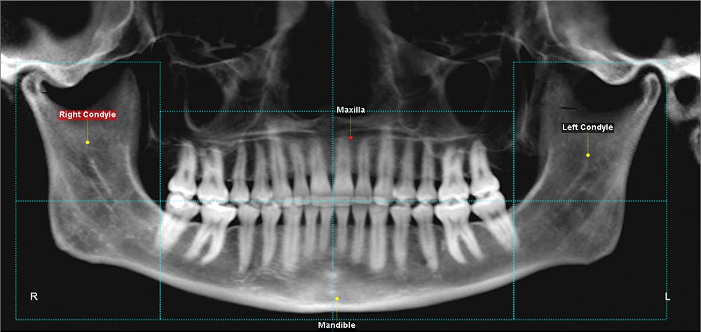
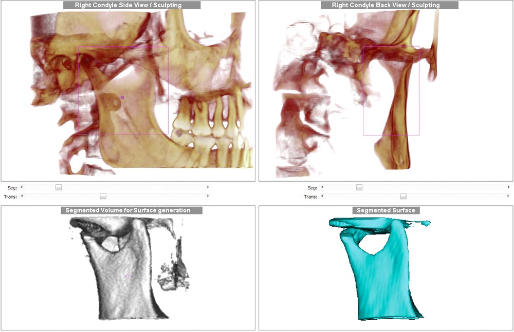
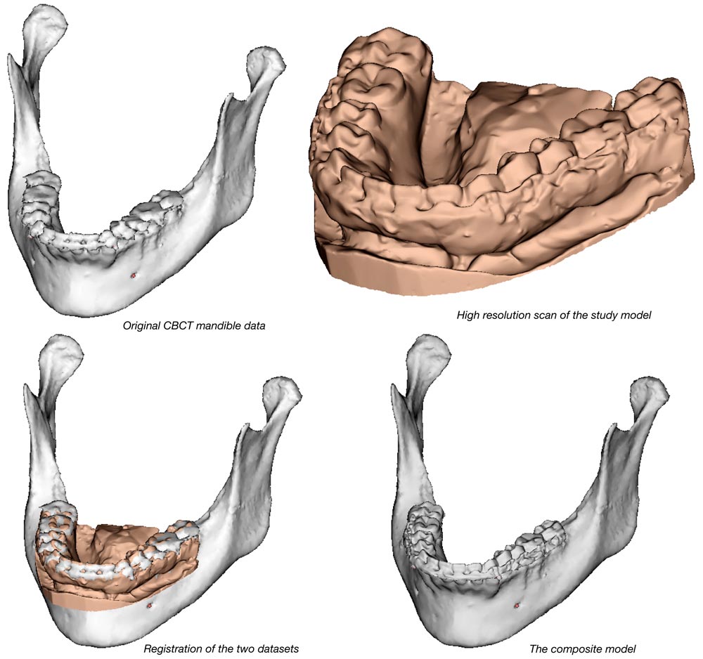
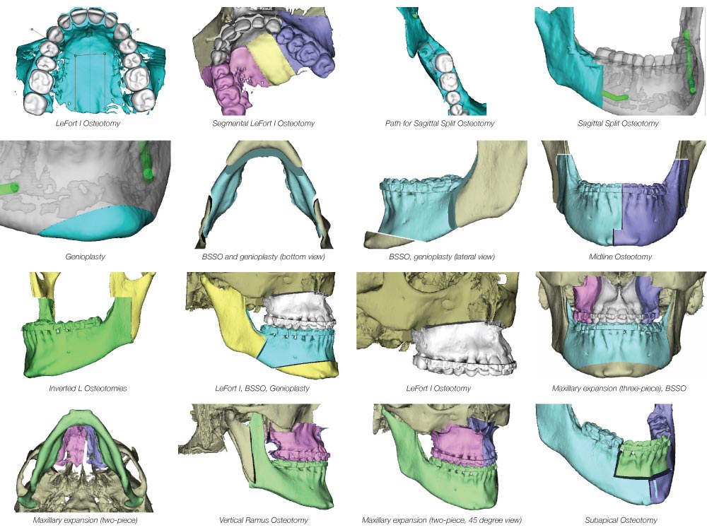
3D Surgery is a comprehensive case planning and presentation tool that animates the patient’s skeletal and facial changes in real time, and outputs to a precise surgical guide. All you need is a 3D DICOM dataset, virtual models and optional facial photo. Use data from cone beam CT, spiral CT, and other sources. Combine with intraoral scans or electronic models for accurate virtual model surgery. Along with the other wonderful features of Dolphin, this is why Dolphin 3D is used worldwide.

Simple to Learn, Easy to Use

Begin with a quality 3D cone beam or spiral CT image. Then, simply follow the step-by-step Segmentation Wizard to create the proper virtual surgery patient. If you utilize a 3D facial camera, Dolphin can process it; if not, Dolphin 3D Surgery can take full advantage of your clinical (2D) photographs.

Treat: Comprehensive Surgical Planning

The heart of Dolphin’s 3D Surgery module is a very powerful and comprehensive surgical planning tool called Treat. You can plan from lateral, front or submento-vertex (SMV) views. You have access to many useful clinical and numeric planning tools designed specifically by and for surgeons, technicians and researchers. Use these tools to produce quick workups; interactive discussion with other specialists; or a detailed plan with splints for the operating room.

Present: Animated Treatment Presentation

The Present tool uses the simulated treatment from Treat in an animated sequence of pre-/post-operation configurations in all three dimensions. This allows you to carefully study detailed treatment nuances; demonstrate your plan to the patient; or conduct case presentation to your surgical team.

Splint: Customizable Surgical Guide Fabrication

Splint tool allows you to select the width, thickness and other parameters of the splint based on your treatment plans. Create an intermediate splint to position the maxilla or mandible based on your preferred order of operation. And then create a final splint. All splint data files are generated in industry standard .STL format, for physical printing at the lab of your choice, or even on your own in-office 3D printer.

Easy steps to create 3D surgical work-up

Step 1: Setup – Map out the general area of the main structures: Proximal segments, maxilla and mandible.


Step 2: Crop – Precisely segment the for maxilla and mandible.


Step 3: Clean-up and merge – Sculpt away any excess tissue from around the maxilla and mandible, and replace low-resolution CBCT teeth with auto-merged high-resolution, digital study model teeth.


Step 4: Osteotomy – Perform the custom osteomy specific to the patient/case.


Step 5: Landmark – Digitize critical landmarks, including any 3D cephalometric analysis landmarks.

Requirements - Specifications